Pages


Sabtu, 22 Desember 2012

Viral hemorrhagic fevers

2 komentar
Viral hemorrhagic fevers

Definition:
Viral hemorrhagic fevers

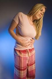
Viral hemorrhagic (hem-uh-RAJ-ik) fevers are infectious diseases that interfere with the blood's natural ability to clot. These diseases can also damage the walls of tiny blood vessels, making them leaky. The internal bleeding that results can range from relatively minor to life-threatening.
Some viral hemorrhagic fevers include:
  • Dengue
  • Ebola
  • Lassa
  • Marburg
  • Yellow fever
These diseases most commonly occur in tropical areas of the world. When viral hemorrhagic fevers occur in the United States, they're usually found in people who've recently traveled internationally.
Viral hemorrhagic fevers are spread by contact with infected animals, people or insects. No current treatment can cure viral hemorrhagic fevers, and immunizations exist for only a few types. Until additional vaccines are developed, the best approach is prevention.

Symptoms:

Signs and symptoms of viral hemorrhagic fevers vary by disease. In general, initial symptoms may include:
  • High fever
  • Fatigue
  • Dizziness
  • Muscle aches
  • Weakness
Symptoms can become life-threatening
Severe cases of some types of viral hemorrhagic fevers may cause bleeding:
  • Under the skin
  • In internal organs
  • From the mouth, eyes or ears
Other signs and symptoms of severe infections can include:
  • Shock
  • Nervous system malfunctions
  • Coma
  • Delirium
  • Seizures
  • Kidney failure
When to see a doctor
The best time to see a doctor is before you travel to a developing country to ensure you've received any available vaccinations and pre-travel advice for staying healthy. If you develop signs and symptoms once you return home, consider consulting a doctor who focuses on international medicine or infectious diseases. A specialist may be able to recognize and treat your illness faster. Be sure to let your doctor know what areas you've visited.

Causes:

The viruses that cause viral hemorrhagic fevers live naturally in a variety of animal and insect hosts — most commonly mosquitoes, ticks, rodents or bats.
Each of these hosts typically lives in a specific geographic area, so each particular disease usually occurs only where that virus's host normally lives. Some viral hemorrhagic fevers also can be transmitted from person to person.

How is it transmitted?
The route of transmission varies by specific virus. Some viral hemorrhagic fevers are spread by mosquito or tick bites. Others are transmitted by contact with infected blood or semen. A few varieties are breathed in if you're around infected rat feces or urine.

If you travel to an area where a particular hemorrhagic fever is common, you may become infected there and then develop symptoms after you return home

Complications:

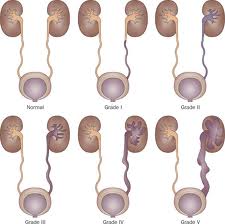
Viral hemorrhagic fevers can damage your:
  • Brain
  • Eyes
  • Heart
  • Kidneys
  • Liver
  • Lungs
  • Spleen
In some cases, the damage is severe enough to cause death.

Treatments and drugs:

Medications
While no specific treatment exists for most viral hemorrhagic fevers, the antiviral drug ribavirin (Virazole, Rebetol) may help shorten the course of infection and prevent complications in some cases.

Therapy
Supportive care is essential. To prevent dehydration, you may need fluids to help maintain your balance of electrolytes — minerals that are critical to nerve and muscle function.

Surgical and other procedures
Some people may benefit from kidney dialysis, an artificial way of cleaning wastes from your blood when your kidneys fail.
Read more...

Viral gastroenteritis (stomach flu)

2 komentar
Viral gastroenteritis (stomach flu)

Definition:
Viral gastroenteritis (stomach flu)
Viral gastroenteritis is an intestinal infection marked by watery diarrhea, abdominal cramps, nausea or vomiting, and sometimes fever.

The most common way to develop viral gastroenteritis — often called stomach flu — is through contact with an infected person or ingestion of contaminated food or water. If you're otherwise healthy, you'll likely recover without complications. But for infants, older adults and people with compromised immune systems, viral gastroenteritis can be deadly.

There's no effective treatment for viral gastroenteritis, so prevention is key. In addition to avoiding food and water that may be contaminated, thorough and frequent hand-washing is your best defense.

Symptoms  :

Although it's commonly called stomach flu, gastroenteritis isn't the same as influenza. Real flu (influenza) affects your respiratory system — your nose, throat and lungs. Gastroenteritis, on the other hand, attacks your intestines, causing signs and symptoms such as:
  • Watery, usually nonbloody diarrhea — bloody diarrhea usually means you have a different, more severe infection
  • Abdominal cramps and pain
  • Nausea, vomiting or both
  • Occasional muscle aches or headache
  • Low-grade fever
Depending on the cause, viral gastroenteritis symptoms may appear within one to three days after you're infected and can range from mild to severe. Symptoms usually last just a day or two, but occasionally they may persist as long as 10 days.

Because the symptoms are similar, it's easy to confuse viral diarrhea with diarrhea caused by bacteria such as salmonella and E. coli or parasites such as giardia.

When to see a doctor
If you're an adult, call your doctor if:
  • You're not able to keep liquids down for 24 hours
  • You've been vomiting for more than two days
  • You're vomiting blood
  • You're dehydrated — signs of dehydration include excessive thirst, dry mouth, deep yellow urine or little or no urine, and severe weakness, dizziness or lightheadedness
  • You notice blood in your bowel movements
  • You have a fever above 104 F (40 C)
For infants and children
See your doctor right away if your child:
  • Has a fever of 102 F (38.9 C) or higher
  • Seems lethargic or very irritable
  • Is in a lot of discomfort or pain
  • Has bloody diarrhea
  • Seems dehydrated — watch for signs of dehydration in sick infants and children by comparing how much they drink and urinate with how much is normal for them
If you have an infant, remember that while spitting up may be an everyday occurrence for your baby, vomiting is not. Babies vomit for a variety of reasons, many of which may require medical attention.
Call your baby's doctor right away if your baby:
  • Has vomiting that lasts more than several hours
  • Hasn't had a wet diaper in six hours
  • Has bloody stools or severe diarrhea
  • Has a sunken fontanel — the soft spot on the top of your baby's head
  • Has a dry mouth or cries without tears
  • Is unusually sleepy, drowsy or unresponsive
Causes:

You're most likely to contract viral gastroenteritis when you eat or drink contaminated food or water, or if you share utensils, towels or food with someone who's infected.
Viruses that can cause gastroenteritis include:
  • Rotavirus. Children are usually infected when they put their fingers or other objects contaminated with the virus into their mouths. Adults infected with rotavirus may not have symptoms, but can still spread the illness — of particular concern in institutional settings because infected adults unknowingly can pass the virus along to others. Some people, particularly those in institutional settings, may spread the virus even though they don't have any symptoms of illness themselves. A vaccine against rotaviral gastroenteritis is available in some countries, including the United States, and appears to be effective in preventing severe symptoms.
  • Noroviruses. Both children and adults are affected by noroviruses. Norovirus infection can sweep through families and communities. It's especially likely to spread among people in confined spaces. In most cases you pick up the virus from contaminated food or water, although person-to-person transmission also is possible.
Some shellfish, especially raw or undercooked oysters, also can make you sick. Contaminated drinking water is another cause of viral diarrhea. But in many cases, the virus is passed through the fecal-oral route — that is, someone with the virus handles food you eat without washing his or her hands after using the toilet.

Complications:

The main complication of viral gastroenteritis is dehydration — a severe loss of water and essential salts and minerals. If you're healthy and drink enough to replace fluids you lose from vomiting and diarrhea, dehydration shouldn't be a problem.
Infants, older adults and people with suppressed immune systems may become severely dehydrated when they lose more fluids than they can replace. If that's the case, hospitalization might be needed so that lost fluids can be replaced intravenously. Rarely, dehydration can be fatal.

Treatments and drugs:
 
 There's often no specific medical treatment for viral gastroenteritis. Antibiotics aren't effective against viruses, and overusing them can contribute to the development of antibiotic-resistant strains of bacteria. Treatment consists of self-care measures.
Read more...

Trench mouth

1 komentar
Trench mouth

Definition  :
Trench mouth

Trench mouth is a severe form of gingivitis that causes painful, infected, bleeding gums and ulcerations. Although trench mouth is rare today in developed nations, it's common in developing countries that have poor nutrition and poor living conditions.

Trench mouth is formally known as Vincent's stomatitis, acute necrotizing ulcerative gingivitis (ANUG) and necrotizing ulcerative gingivitis (NUG). Trench mouth earned its nickname because of its prevalence among soldiers who were stuck in the trenches during World War I without the means to take care of their teeth properly.

Symptoms:

Signs and symptoms of trench mouth can include:
  • Severe gum pain
  • Bleeding from gums when they're pressed even slightly
  • Red or swollen gums
  • Pain when eating or swallowing
  • A gray film on your gums
  • Crater-like sores (ulcers) between your teeth and on your gums
  • A foul taste in your mouth
  • Bad breath
  • Fever
  • Swollen lymph nodes around your head, neck or jaw
When to see a dentist
Trench mouth symptoms can develop quickly. See your dentist immediately if you develop any symptoms. Often these may be symptoms of a gum problem other than trench mouth, such as gingivitis or periodontitis. But all forms of gum disease can be serious, and most tend to get worse without treatment. The sooner you seek care, the better your chance of returning your gums to a healthy state and preventing loss of teeth and destruction of bone or other tissue.

Causes:

Your mouth naturally contains microorganisms, including fungi, viruses and bacteria. In trench mouth, though, harmful bacteria grow out of control, causing infection of your gums. This infection can damage or destroy the delicate gum tissue (gingiva) that surrounds and supports your teeth. Large ulcers, often filled with bacteria, food debris and decaying tissue, may form on your gums, leading to severe pain, bad breath and a foul taste in your mouth.

Precisely how these bacteria destroy gum tissue isn't known. But it's thought that enzymes and toxins produced by the bacteria play a role.

Complications:
 
Complications and problems that trench mouth may cause or be associated with include:
  • Trouble swallowing
  • Trouble eating
  • Pain when brushing teeth
  • Destruction of gum tissue
  • Tooth loss
  • Progression into advanced oral diseases that can severely damage bone and gum tissue
Treatments and drugs:

Treatment of trench mouth is generally highly effective, and complete healing often occurs in just a couple of weeks. However, healing may take longer if your immune system is compromised, such as by HIV/AIDS.

Medications
Because trench mouth involves an overgrowth of bacteria, antibiotics are often prescribed to eradicate the bacteria and prevent infection from spreading. You may also need over-the-counter or prescription pain relievers. Getting pain under control is important so that you can eat properly and resume good dental care habits, such as brushing your teeth. Your dentist may also recommend a pain reliever that you can apply directly to your gums (topical anesthetic).

Cleaning your teeth and gums
Treatment also includes a thorough but gentle cleaning of your teeth and gums. Your dentist removes any dead gum tissue (debridement) to help reduce pain. Your mouth may be rinsed with an antiseptic solution. When your gums are less tender, you'll undergo a type of tooth cleaning called scaling and root planing. This procedure removes plaque and tartar from beneath your gumline and smooths any roughened surfaces of your teeth.
Right after cleaning, your gums will be quite tender. Your dentist will probably advise you to rinse your mouth with a hydrogen peroxide mouthwash, salt water or a prescription mouth rinse, in addition to brushing gently with a soft toothbrush. Once your gums begin to heal, brush and floss at least twice a day — preferably after every meal and at bedtime — to prevent future problems.

When surgery is necessary
Although your gums are likely to heal and return to their normal shape with professional cleaning and proper home care, you may need surgery to help repair them if you have extensive damage.
Read more...

Vesicoureteral reflux

2 komentar
Vesicoureteral reflux

Definition:
Vesicoureteral reflux

Vesicoureteral (ves-ih-koe-yoo-REE-ter-ul) reflux is the abnormal flow of urine from your bladder back up the tubes (ureters) that connect your kidneys to your bladder. Normally, urine flows only down from your kidneys to your bladder.

Vesicoureteral reflux is usually diagnosed in infants and children. The disorder increases the risk of urinary tract infections, which, if left untreated, can lead to kidney damage.

Vesicoureteral reflux can be primary or secondary. Children with primary vesicoureteral reflux are born with a defect in the valve that normally prevents urine from flowing backward from the bladder into the ureters. Secondary vesicoureteral reflux is due to a urinary tract malfunction, often caused by infection.

Children may outgrow primary vesicoureteral reflux. Treatment, which includes medication or surgery, aims at preventing kidney damage.

Symptoms:

A urinary tract infection (UTI) is the most common indication of vesicoureteral reflux. A UTI doesn't always cause noticeable signs and symptoms, though most people have some. These signs and symptoms can include:
  • A strong, persistent urge to urinate
  • A burning sensation when urinating
  • Passing frequent, small amounts of urine
  • Blood in the urine (hematuria) or cloudy, strong-smelling urine
  • Fever
  • Abdominal or flank pain
  • Hesitancy to urinate or holding urine to avoid the burning sensation
A UTI may be difficult to diagnose in children, who may have only nonspecific signs and symptoms. Signs and symptoms in infants with a UTI may also include:
  • Diarrhea
  • Lack of appetite
  • An unexplained fever
  • Irritability
As your child gets older, untreated vesicoureteral reflux can lead to other signs and symptoms, including:
  • Bed-wetting
  • High blood pressure
  • Protein in urine
  • Kidney failure
Another indication of vesicoureteral reflux, which may be detected before birth by sonogram, is swelling of the kidneys or the urine-collecting structures of one or both kidneys (hydronephrosis) in the fetus, caused by the backup of urine into the kidneys.
When to see a doctor
Contact your doctor right away if your child develops any of the signs or symptoms of a UTI, such as:
  • A strong, persistent urge to urinate
  • A burning sensation when urinating
  • Abdominal or flank pain
  • A hesitancy to urinate
Call your doctor about fever if your child:
  • Is less than 3 months old and has a rectal temperature of 100.4 F (38 C) or higher
  • Is 3 months or older and has a fever of 104 F (40 C)
In addition, call your doctor immediately if your infant has the following signs or symptoms:
  • Changes in appetite. If your baby refuses several feedings in a row or eats poorly, contact the doctor.
  • Changes in mood. If your baby is lethargic or unusually difficult to rouse, tell the doctor right away. Also let the doctor know if your baby is persistently irritable or has periods of inconsolable crying.
  • Diarrhea. Contact the doctor if your baby's stools are especially loose or watery.
  • Vomiting. Occasional spitting up is normal. Contact the doctor if your baby spits up large portions of multiple feedings or vomits forcefully after feedings.
Causes:

Your urinary system includes your kidneys, ureters, bladder and urethra. All play a role in removing waste products from your body.
The kidneys, a pair of bean-shaped organs at the back of your upper abdomen, filter waste, water and electrolytes — minerals, such as sodium, calcium and potassium, that help maintain the balance of fluids in your body — from your blood. Tubes called ureters carry urine from your kidneys down to your bladder, where it is stored until it exits the body through another tube (the urethra) during urination.

Vesicoureteral reflux can develop in two forms, primary and secondary:
  • Primary vesicoureteral reflux. The cause of this more common form is a defect that's present before birth (congenital). The defect is in the functional valve between the bladder and a ureter that normally closes to prevent urine from flowing backward. As the child grows, the ureters lengthen and straighten, which may improve valve function and eventually resolve the reflux. This type of vesicoureteral reflux tends to run in families, which indicates that it may be genetic, but the exact cause of the defect is unknown.
  • Secondary vesicoureteral reflux. The cause of this form is a blockage or malfunction in the urinary system. The blockage most commonly results from recurrent UTIs, which may cause swelling of a ureter
Complications:

Kidney damage is the primary concern with vesicoureteral reflux. The more severe the reflux, the more serious the complications are likely to be. Complications may include:
  • Kidney (renal) scarring. Untreated UTIs can lead to scarring, also known as reflux nephropathy, which is permanent damage to kidney tissue. A backup of urine exposes the kidneys to higher than normal pressure, which can lead to scarring over time. Extensive scarring may lead to high blood pressure and kidney failure.
  • High blood pressure (hypertension). Because the kidneys remove waste from the bloodstream, damage to your kidneys and the resultant buildup of wastes can raise your blood pressure.
  • Kidney failure. Scarring can cause a loss of function in the filtering part of the kidney. This may lead to kidney failure, which can occur quickly (acute) or it may develop over time (chronic). In either case, dialysis or a kidney transplant may be necessary. Dialysis is an artificial means of removing extra fluids and waste from your blood.
Treatments and drugs:

Treatment options for vesicoureteral reflux depend on the severity of the condition. Children with mild cases of primary vesicoureteral reflux may eventually outgrow the disorder. In this case, your doctor will likely recommend a wait-and-see approach. During this time, it will be important for you to be watchful for potential UTIs and to seek prompt treatment.

Children with moderate to severe primary vesicoureteral reflux have two treatment options: medication and surgery. Using medication is more common, with surgery usually reserved for those children for whom antibiotics aren't successful.
However, surgery may be a first line therapy for grades IV and V or for families who prefer a quicker, more definitive treatment than medication.

Medications
UTIs require prompt treatment with antibiotics to keep the infection from moving to the kidneys. Doctors may also use antibiotics to prevent UTIs, usually at about half the dose for treating an infection.

Commonly used antibiotics for prevention include the combination drug trimethoprim-sulfamethoxazole (Bactrim, Septra), trimethoprim (Primsol) and nitrofurantoin (Furadantin, Macrobid, Macrodantin). Some people may be allergic to one or more of these medications, preventing their use. Possible side effects of long-term use of these drugs include:
  • Nausea and vomiting
  • Abdominal pain
  • Increased antibiotic resistance, in which the infection no longer responds to antibiotics and becomes more difficult to treat
A child being treated with medication needs to be monitored for as long as he or she is taking antibiotics. This includes periodic physical exams and urine tests to detect breakthrough infections — UTIs that occur despite the antibiotic treatment — and occasional radiographic scans of the bladder and kidneys to determine if your child has outgrown vesicoureteral reflux.

Surgery
Surgery for vesicoureteral reflux repairs the defect in the functional valve between the bladder and each affected ureter that keeps it from closing and preventing urine from flowing backward. There are two methods of surgical repair:
  • Open surgery. Performed using general anesthesia, this surgery requires an incision in the lower abdomen through which the surgeon repairs the malformation that's causing the problem. This type of surgery usually requires a few days' stay in the hospital, during which a catheter is kept in place to drain your child's bladder. Risks include infection, blood clots and bleeding.
  • Endoscopic surgery. In this procedure, the doctor inserts a lighted tube (cystoscope) through the urethra to see inside your child's bladder, then injects a bulking agent around the opening of the affected ureter to try to strengthen the valve's ability to close properly. This method is minimally invasive compared with open surgery and presents fewer risks, though it may not be as effective. This procedure also requires general anesthesia, but generally can be performed as outpatient surgery.
Read more...

Benign paroxysmal positional vertigo (BPPV)

1 komentar
Benign paroxysmal positional vertigo (BPPV)

Definition:
Benign paroxysmal positional vertigo (BPPV)

Benign paroxysmal positional vertigo (BPPV) is one of the most common causes of vertigo — the sudden sensation that you're spinning or that the inside of your head is spinning.
Benign paroxysmal positional vertigo is characterized by brief episodes of mild to intense dizziness. Symptoms of benign paroxysmal positional vertigo are triggered by specific changes in the position of your head, such as tipping your head up or down, and by lying down, turning over or sitting up in bed. You may also feel out of balance when standing or walking.

Although benign paroxysmal positional vertigo can be a bothersome problem, it's rarely serious except when it increases the chance of falls. You can receive effective treatment for benign paroxysmal positional vertigo during a doctor's office visit.

Symptoms:

The signs and symptoms of benign paroxysmal positional vertigo (BPPV) may include:
  • Dizziness
  • A sense that you or your surroundings are spinning or moving (vertigo)
  • Lightheadedness
  • Unsteadiness
  • A loss of balance
  • Blurred vision associated with the sensation of vertigo
  • Nausea
  • Vomiting
The signs and symptoms of BPPV can come and go, with symptoms commonly lasting less than one minute. Episodes of benign paroxysmal positional vertigo and other forms of vertigo can disappear for some time and then recur.
Activities that bring about the signs and symptoms of BPPV can vary from person to person, but are almost always brought on by a change in the position of your head. Abnormal rhythmic eye movements (nystagmus) usually accompany the symptoms of benign paroxysmal positional vertigo. Although rare, it's possible to have BPPV in both ears (bilateral BPPV).

When to see a doctor
Generally, see your doctor if you experience any unexplained dizziness or vertigo that recurs periodically for more than one week.

Seek emergency care
Although it's uncommon for dizziness to signal a serious illness, see your doctor immediately if you experience dizziness or vertigo along with any of the following:
  • A new, different or severe headache
  • A fever of 101 F (38 C) or higher
  • Double vision or loss of vision
  • Hearing loss
  • Trouble speaking
  • Leg or arm weakness
  • Loss of consciousness
  • Falling or difficulty walking
  • Numbness or tingling
  • Chest pain, or rapid or slow heart rate
The signs and symptoms listed above may signal a more serious problem, such as stroke or a cardiac condition

Causes:

About half the time, doctors can't find a specific cause for BPPV.
When a cause can be determined, BPPV is often associated with a minor to severe blow to your head. Less common causes of BPPV include disorders that damage your inner ear or, rarely, damage that occurs during ear surgery or during prolonged positioning on your back. BPPV also has been associated with migraines.

The ear's role
Inside your ear is a tiny organ called the vestibular labyrinth. It includes three loop-shaped structures (semicircular canals) that contain fluid and fine, hair-like sensors that monitor the rotation of your head.

Other structures (otolith organs) in your ear monitor movements of your head — up and down, right and left, back and forth — and your head's position related to gravity. These otolith organs — the utricle and saccule — contain crystals that make you sensitive to gravity.

For a variety of reasons, these crystals can become dislodged. When they become dislodged, they can move into one of the semicircular canals — especially while you're lying down. This causes the semicircular canal to become sensitive to head position changes it would normally not respond to. As a result, you feel dizzy.

Complications:

 Although benign paroxysmal positional vertigo (BPPV) is uncomfortable, it rarely causes complications. In rare cases, if severe, persistent BPPV causes you to vomit frequently, you may be at risk of dehydration. The dizziness of BPPV can put you at greater risk of falling.

Treatments and drugs:

To help relieve benign paroxysmal positional vertigo (BPPV), your doctor, audiologist or physical therapist may treat you with a series of movements known as the canalith repositioning procedure.

Canalith repositioning
Performed in your doctor's office, the canalith repositioning procedure consists of several simple and slow maneuvers for positioning your head. The goal is to move particles from the fluid-filled semicircular canals of your inner ear into a tiny bag-like open area (vestibule) that houses one of the otolith organs (utricle) in your ear where these particles don't cause trouble and are more easily resorbed. Each position is held for about 30 seconds after any symptoms or abnormal eye movements stop. This procedure is usually effective after one or two treatments.

After the procedure, you must avoid lying flat or placing the treated ear below shoulder level for the rest of that day. For the first night following the procedure, elevate your head on a few pillows when you sleep. This allows time for the particles floating in your labyrinth to settle into your vestibule and be resorbed by the fluids in your inner ear.

On the morning after your in-office procedure, your restrictions will be lifted and you'll begin self-care as directed by your doctor. Your doctor likely will have taught you how to perform the canalith repositioning procedure on yourself so that you can do it at home before returning to the office for a recheck.

Surgical alternative
In very rare situations in which the canalith repositioning procedure isn't effective, your doctor may recommend a surgical procedure in which a bone plug is used to block the portion of your inner ear that's causing dizziness. The plug prevents the semicircular canal in your ear from being able to respond to particle movements or head movements in general. This success rate for canal plugging surgery is greater than 90 percent.
Read more...

Ventricular septal defect (VSD)

3 komentar
Ventricular septal defect (VSD)

Definition:
Ventricular septal defect

A ventricular septal defect (VSD), also called a hole in the heart, is a common heart defect that's present at birth (congenital). The defect involves an opening (hole) in the heart forming between the heart's lower chambers, allowing oxygen-rich and oxygen-poor blood to mix.

A baby with a small ventricular septal defect may have no problems. A baby with a larger ventricular septal defect or associated heart defects may have a telltale bluish tint to the skin (cyanosis) — due to oxygen-poor blood — often most visible in the lips and fingernails. Ventricular septal defects are sometimes not diagnosed until adulthood.

Fortunately, ventricular septal defect is treatable. Many small ventricular septal defects often close on their own or don't cause problems. Larger ventricular septal defects need surgical repair early in life to prevent complications. Some smaller ventricular septal defects are closed to prevent complications related to their location, such as damage to heart valves. Many people with small ventricular septal defects have normal, productive lives with few related problems.

Symptoms:

Signs and symptoms of serious heart defects often appear during the first few days, weeks or months of a child's life.
Ventricular septal defect symptoms in a baby may include:
  • A bluish tint to the skin, lips and fingernails (cyanosis)
  • Poor eating, failure to thrive
  • Fast breathing or breathlessness
  • Easy tiring
  • Swelling of legs, feet or abdomen
  • Rapid heart rate
Although these signs can be caused by other conditions, they may be due to a congenital heart defect.
You and your doctor may not notice signs of a ventricular septal defect at birth. If the defect is small, symptoms may not appear until later in childhood — if ever. Signs and symptoms vary depending on the size of the hole. Your doctor may first suspect a heart defect during a regular checkup while listening to your baby's heart with a stethoscope.

Sometimes a ventricular septal defect isn't detected until a person reaches adulthood. Signs or symptoms can include shortness of breath and a loud heart murmur your doctor can hear when listening to your heart with a stethoscope.

When to see a doctor
Call your doctor if your baby or child:
  • Tires easily when eating or playing
  • Is not gaining weight
  • Becomes breathless when eating or crying
  • Has a bluish tint to his or her skin, especially around the fingernails and lips
  • Breathes rapidly or is short of breath
Call your doctor if you develop:
  • Shortness of breath when you exert yourself or when you lie down
  • Rapid or irregular heartbeat
  • Fatigue and weakness
  • Swelling (edema) in your legs, ankles and feet
Causes:

Heart defects that are present at birth (congenital) arise from problems early in the heart's development, but there's often no clear cause. Genetics and environmental factors probably play a role.
A ventricular septal defect occurs when the septum, the muscular wall separating the heart into left and right sides, fails to form fully between the lower chambers of the heart (ventricles) during fetal development. This leaves an opening that allows mixing of oxygenated blood and deoxygenated blood, meaning the heart has to work harder to provide enough oxygen to your body's tissues.

If a ventricular septal defect is large, blood overfills the lungs and overworks the heart. If left untreated, the blood pressure in the lungs goes up (pulmonary hypertension) and the ventricles enlarge and no longer work efficiently. Ultimately, this can lead to irreversible damage to the lung arteries and to heart failure. In contrast, small ventricular septal defects don't usually cause any problems, except for a loud heart murmur that doctors may note during physical exams.

Complications:

A small ventricular septal defect may never cause any problems. Larger defects can cause a range of disabilities — from mild to life-threatening. Treatment can prevent many complications.

Eisenmenger's syndrome
If a large ventricular septal defect goes untreated, increased blood flow to the lungs causes high blood pressure in the lung arteries (pulmonary hypertension). Over time, permanent damage to the lung arteries develops and the pulmonary hypertension can become irreversible.

This complication, called Eisenmenger's syndrome, usually develops in early childhood. In people with Eisenmenger's syndrome, a significant portion of blood flows through the ventricular septal defect from the right ventricle to the left and bypasses the lungs.

This means deoxygenated blood is pumped to the body and leads to a bluish discoloration of the lips, fingers and toes (cyanosis) and other complications. Once a person has Eisenmenger's syndrome, it's too late to surgically repair the hole because irreversible damage to the lung arteries has already occurred.

Other complications
Other complications may include:
  • Heart failure. The increased blood flow through the heart due to a ventricular septal defect can also lead to heart failure, a chronic condition in which the heart can't pump effectively.
  • Endocarditis. People with a ventricular septal defect are at increased risk of an infection of the heart (endocarditis).
  • Stroke. People with large defects, especially occurring with Eisenmenger's syndrome, are at risk of a stroke due to a blood clot passing through the hole in the heart and going to the brain.
  • Other heart problems. Ventricular septal defects can also lead to abnormal heart rhythms and valve problems.
Ventricular septal defect and pregnancy
Becoming pregnant is often a concern for women born with a heart defect. Having a repaired ventricular septal defect without any complications or having a very small defect doesn't pose any additional risk in pregnancy. However, having an unrepaired larger defect, heart failure, cyanosis or other heart defects poses a high risk to both mother and fetus. Women with Eisenmenger's syndrome are at the highest risk of complications.

Doctors strongly advise these women not to become pregnant.
Any woman with a congenital heart defect, repaired or not, who is considering pregnancy should talk beforehand with a doctor who specializes in the diagnosis and treatment of heart conditions (cardiologist). This is especially important if you're taking medications. It's also important to see both an obstetrician and a cardiologist during pregnancy.

Treatments and drugs:

Many babies born with a small ventricular septal defect won't ever need to have the defect surgically closed. After birth, your doctor may want to observe your baby and treat any symptoms while waiting to see if the defect will close on its own.

Children and adults who have a ventricular septal defect that is large or is causing significant symptoms usually need surgery to close the defect. If your baby has a ventricular septal defect that needs surgical repair, the procedure will likely be scheduled in your baby's first year of life.
Medications
Medications for ventricular septal defect may include those to:
  • Keep the heartbeat regular. Examples include beta blockers (Lopressor, Inderal, others) and digoxin (Lanoxin, Lanoxicaps, Lanoxin Pediatric).
  • Increase the strength of the heart's contractions. Examples include digoxin (Lanoxin).
  • Decrease the amount of fluid in circulation. Doing so reduces the volume of blood that must be pumped. These medications, called diuretics, include furosemide (Lasix).
Procedures
Surgical treatment for ventricular septal defects involves plugging or patching the abnormal opening between the ventricles. Two approaches are used:
  • Surgical repair. This is the procedure of choice in most cases. Surgical repair of a ventricular septal defect usually involves open-heart surgery, which is done under general anesthesia. The surgery requires a heart-lung machine and an incision in the chest. The doctor uses patches or stitches to close the hole.
  • Catheter procedure. This method may be used to close some ventricular septal defects. Patching during catheterization doesn't require opening the chest. Rather than opening the chest, the doctor inserts a thin tube (catheter) into a blood vessel in the groin and guides it to the heart. The doctor then uses a small mesh patch or plug to close the hole.
  • Hybrid procedure. A hybrid procedure uses surgical and catheter-based techniques. Access to the heart is usually through a small incision and the procedure may be performed without stopping the heart and using the heart-lung machine. A plug is delivered to close the VSD via a catheter placed through the small hole that the surgeon created. Recovery from this procedure is quicker than with standard surgery.
After repair, your doctor will schedule regular medical follow-up to ensure that the ventricular septal defect remains closed. Depending on the size of the ventricular septal defect and the presence or absence of any other problems, your doctor will tell you how frequently you or your child will need to be seen.
Surgery to close a ventricular septal defect generally has excellent long-term results.

Preventive antibiotics
If you've been told in the past that you or your child needs to take antibiotics before dental or medical procedures to reduce the risk of infective endocarditis, talk with your doctor. Endocarditis is much more likely to occur from exposure to random germs than from a typical dental exam or surgery. Current guidelines recommend preventive antibiotic treatment only for those people at highest risk of serious complications from infective endocarditis.
Your doctor may still recommend preventive antibiotics if you:
  • Have other heart conditions or artificial valves
  • Have a large ventricular septal defect that's causing a low blood oxygen level
  • Have had a repair with artificial (prosthetic) material
For most people with a ventricular septal defect, practicing good oral hygiene and getting regular dental checkups is the most effective strategy for preventing endocarditis.
Read more...

Endometrial cancer

2 komentar
Endometrial cancer

Definition:
Endometrial cancer

Endometrial cancer is a type of cancer that begins in the uterus. The uterus is the hollow, pear-shaped pelvic organ in women where fetal development occurs.

Endometrial cancer begins in the layer of cells that form the lining (endometrium) of the uterus. Endometrial cancer is sometimes called uterine cancer. Other types of cancer can form in the uterus, but they are much less common than endometrial cancer.

Endometrial cancer is often detected at an early stage because it frequently produces abnormal vaginal bleeding, which prompts women to see their doctors. If endometrial cancer is discovered early, removing the uterus surgically often eliminates all of the cancer.

Symptoms:

Signs and symptoms of endometrial cancer may include:
  • Vaginal bleeding after menopause
  • Prolonged periods or bleeding between periods
  • An abnormal, watery or blood-tinged discharge from your vagina
  • Pelvic pain
  • Pain during intercourse
When to see a doctor
Make an appointment with your doctor if you experience any signs or symptoms that worry you, such as vaginal bleeding or discharge not related to your periods, pelvic pain, or pain during intercourse.

Causes:

 Doctors don't know what causes endometrial cancer. What's known is that something occurs to create a genetic mutation within cells in the endometrium — the lining of the uterus. The genetic mutation turns normal, healthy cells into abnormal cells.

Healthy cells grow and multiply at a set rate, eventually dying at a set time. Abnormal cells grow and multiply out of control, and they don't die at a set time. The accumulating abnormal cells form a mass (tumor). Cancer cells invade nearby tissues and can separate from an initial tumor to spread elsewhere in the body (metastasize).

Complications:

 Endometrial cancer can spread to other parts of your body, making it more difficult to treat successfully. Endometrial cancer that spreads (metastasizes) most often travels to the lungs.

Treatments and drugs:

Your options for treating your endometrial cancer will depend on the characteristics of your cancer, such as the stage, your general health and your preferences.

Surgery
Surgery to remove the uterus is recommended for most women with endometrial cancer. Most women with endometrial cancer undergo a procedure to remove the uterus (hysterectomy), as well as to remove the fallopian tubes and ovaries (salpingo-oophorectomy). A hysterectomy makes it impossible for you to have children in the future. Also, once your ovaries are removed, you'll experience menopause, if you haven't already.
During surgery, your surgeon will also inspect the areas around your uterus to look for signs that cancer has spread. Your surgeon may also remove lymph nodes for testing. This helps determine your cancer's stage.

Radiation
Radiation therapy uses powerful energy beams, such as X-rays, to kill cancer cells. In some instances, your doctor may recommend radiation to reduce your risk of a cancer recurrence after surgery. If you aren't healthy enough to undergo surgery, you may opt for radiation therapy only. In women with advanced endometrial cancer, radiation therapy may help control cancer-related pain.
Radiation therapy can involve:
  • Radiation from a machine outside your body. Called external beam radiation, during this procedure you lie on a table while a machine directs radiation to specific points on your body.
  • Radiation placed inside your body. Internal radiation, or brachytherapy, involves placing a radiation-filled device, such as small seeds, wires or a cylinder, inside your vagina for a short period of time.
Hormone therapy
Hormone therapy involves taking medications that affect hormone levels in the body. Hormone therapy may be an option if you have advanced endometrial cancer that has spread beyond the uterus. Options include:
  • Medications to increase the amount of progesterone in your body. Synthetic progestin, a form of the hormone progesterone, may help stop endometrial cancer cells from growing.
  • Medications to reduce the amount of estrogen in your body. Hormone therapy drugs can help lower the levels of estrogen in your body or make it difficult for your body to use the available estrogen. Endometrial cancer cells that rely on estrogen to help them grow may die in response to these medications.
Chemotherapy
Chemotherapy uses chemicals to kill cancer cells. You may receive one chemotherapy drug, or two or more drugs can be used in combination. You may receive chemotherapy drugs by pill (orally) or through your veins (intravenously). Chemotherapy may be an option for women with advanced endometrial cancer that has spread beyond the uterus. These drugs enter your bloodstream and then travel through your body, killing cancer cells.

Read more...

Chronic hives (urticaria)

3 komentar
Chronic hives (urticaria)

Definition  :
Chronic hives (urticaria)

Chronic hives, also known as urticaria, are batches of raised, red or white itchy welts (wheals) of various sizes that appear and disappear. While most cases of hives go away within a few weeks or less, for some people they are a long-term problem. Chronic hives are defined as hives that last more than six weeks or hives that go away, but recur frequently.

In most cases of chronic hives, a cause is never clearly identified. In some cases, chronic hives may be related to an underlying autoimmune disorder, such as thyroid disease or lupus.
While the underlying cause of chronic hives is usually not identified, treatment can help with symptoms. For many people, antihistamine medications provide the best relief.

Symptoms:

Hives generally:
  • Appear as small round wheals, rings or large patches and may change shape
  • Itch and may be surrounded by a red flare
  • Occur in batches, and often appear on the face or the extremities
Individual hives can last from 30 minutes to 36 hours. As some hives disappear, new hives may develop.

About 40 percent of people with chronic hives also have angioedema. Signs and symptoms of angioedema include large welts or swelling of the skin that may occur around the eyes and lips, hands, feet, genitalia, and inside the throat. Swelling in the throat can obstruct breathing and requires emergency treatment. Angioedema may itch less than hives do, but can cause pain or burning.
Symptoms may not occur all the time. They may come and go with no apparent trigger. For some people, certain conditions — such as heat, exertion or stress — can make symptoms worse.

When to see a doctor
Although chronic hives and angioedema usually aren't life-threatening, they can be debilitating — and in some cases are a sign of an underlying health problem.
See your doctor if you have:
  • Severe hives
  • Hives that don't respond to treatment
  • Hives that continue to appear for several days
Seek emergency care if you:
  • Feel lightheaded
  • Have difficulty breathing
  • Feel your throat is swelling
Causes:

Chronic hives are an inflammation of the skin triggered when certain cells (mast cells) release histamine and other chemicals into your bloodstream, causing small blood vessels to leak. The exact cause of chronic hives isn't well understood — and triggers can be difficult to pinpoint. Chronic hives are thought to be caused by an immune system (autoimmune) disorder and may be linked to another health problem, such as thyroid disease or lupus.

Rarely, a reaction to medication, food, food additives, insects, parasites or infection is identified as an underlying cause of chronic hives. But in most cases, the cause of chronic hives is never identified, even after testing and monitoring symptoms. Heat, cold, pressure, sunlight or other environmental stimuli may worsen chronic hives. Certain pain medications, such as aspirin, ibuprofen (Advil, Motrin, others) and naproxen sodium (Aleve, Anaprox, others), also can worsen chronic hives.

Complications:

Complications of hives and angioedema may include:
  • Itching. Hives and angioedema can cause itching and discomfort.
  • Difficulty breathing. In more-serious cases — when swelling occurs inside your mouth or throat — complications can include difficulty breathing, leading to a loss of consciousness. If you have a swollen throat, seek medical care immediately.

  • Anaphylactic shock (anaphylaxis). This is a serious allergic reaction involving your heart or lungs that can also be associated with hives and angioedema. Your bronchial tubes narrow, it's difficult to breathe, and your blood pressure drops, causing dizziness and perhaps loss of consciousness or even death. Anaphylactic shock occurs rapidly and requires immediate medical care.
Treatments and drugs:

Finding an effective treatment for chronic hives can be challenging. In cases in which a trigger is identified — such as a reaction to a certain food, medication or physical stimulus — treatment includes avoiding the trigger. If your chronic hives are caused by an underlying health condition, they may improve when the underlying condition is treated.

Symptoms can be treated effectively in most people with over-the-counter or prescription medications. Work with your doctor to find the medication — or combination of medications — that works best for you. If the first medication you try doesn't relieve your symptoms, talk to your doctor about trying something else.

Oral antihistamines
These medications block the symptom-producing release of histamine, controlling symptoms for the majority of people with chronic hives — but they do not treat the underlying cause of the rash. Antihistamines are divided into two categories — older, first-generation drugs and newer, second-generation medications. Each category includes nonprescription and prescription drugs. A combination of antihistamines may work best.

Second-generation, newer antihistamines. Your doctor may have you start with newer, nonsedating or low-sedating antihistamines because they are generally as effective and better tolerated than first-generation antihistamines. Examples include:
  • Loratadine (Claritin, Alavert)
  • Fexofenadine (Allegra)
  • Cetirizine (Zyrtec)
  • Levocetirizine (Xyzal)
  • Desloratadine (Clarinex)
First-generation, older antihistamines. If a nonsedating antihistamine doesn't work, your doctor may recommend taking a first-generation antihistamine. These antihistamines can make you drowsy and impair your ability to drive or perform other tasks that require physical coordination. For that reason, your doctor may recommend that you take this type of antihistamine before bedtime and switch to a second-generation drug during the daytime. This class of antihistamines includes:
  • Hydroxyzine (Vistaril)
  • Diphenhydramine (Benadryl)
  • Chlorpheniramine (Chlor-Trimeton)
Check with your doctor before taking any of these medications if you're pregnant or breast-feeding, have a chronic medical condition, or are taking any other medications.

Other medications
If antihistamines alone don't relieve your symptoms, other possible treatments include:
  • H-2 antagonists. These medications, such as cimetidine (Tagamet), ranitidine (Zantac), nizatidine (Axid) and famotidine (Pepcid AC), can be used along with antihistamines. Some common side effects from this class of medications range from gastrointestinal problems to headache.
  • Oral corticosteroids. Oral corticosteroids, such as prednisone, can help lessen swelling, redness and itching — but are usually used only a short term for severe hives or angioedema because they can cause serious side effects. Topical corticosteroids usually aren't effective for chronic hives. Corticosteroids can weaken your immune system, making it easier for you to get an infection or worsening an existing infection you already have.
  • Tricyclic antidepressants. The tricyclic antidepressant doxepin (Zonalon) has antihistamine properties and can help relieve itching. Doxepin may cause dizziness or drowsiness.
Other medications are still being studied to determine whether they may be useful for treating chronic hives. These include:
  • Leukotriene modifiers. Montelukast (Singulair) and zafirlukast (Accolate) are asthma medications that may be helpful when used along with antihistamines. Side effects of these drugs may include behavior and mood changes.
  • Cyclosporine. This immune system suppressant can help with symptoms, but it can cause serious side effects and needs to be monitored carefully. The Food and Drug Administration warns that taking cyclosporine (Gengraf, Neoral, others) puts you at greater risk of opportunistic infections, such as the activation of a previous infection.
  • Omalizumab (Xolair). This medication is normally given by injection to treat allergic asthma. It may help people who have chronic hives caused by an autoimmune response that haven't been helped by antihistamines. Only very small studies have been completed, so more clinical trials are needed.
Read more...

Hives and angioedema

1 komentar
Hives and angioedema

Definition  :
Hives and angioedema

Hives — also known as urticaria (ur-tih-KAR-e-uh) — is a skin reaction that causes raised, red, itchy welts (wheals, or swellings) in sizes ranging from small spots to large blotches several inches in diameter. Individual welts appear and fade as the reaction runs its course. Angioedema is a related type of swelling that affects deeper layers in your skin, often around your eyes and lips.

In most cases, hives and angioedema are harmless and don't leave any lasting marks, even without treatment. The most common treatment for hives and angioedema is antihistamine medications. Serious angioedema can be life-threatening if swelling causes your throat or tongue to block your airway and leads to loss of consciousness.

Symptoms:

Hives
Signs and symptoms of hives include:
  • Raised red or white welts (wheals, or swellings) of various sizes that can cover large areas of skin
  • Welts that resolve while new welts erupt, making it seem as if the condition "moves"
  • Itching, which may be severe
  • Rarely, burning or stinging in the affected area
Hives can be either acute or chronic. Acute hives last from less than one day up to six weeks. Chronic hives last more than six weeks — sometimes for months to years.

Angioedema
Angioedema is a reaction similar to hives that affects deeper layers of your skin, the tissues underneath your skin, and the lining of your throat and intestines. Angioedema often appears around your eyes, cheeks or lips, but can also develop on your hands or feet, or genitals, or inside your throat or bowel.  Angioedema and hives can occur separately or at the same time.
Signs and symptoms of angioedema include:
  • Large, thick, firm welts
  • Swelling of the skin
  • Pain or warmth in the affected areas
  • Difficulty breathing or swallowing, in severe cases
Hereditary angioedema is a rare but more serious inherited (genetic) condition that can cause sudden, severe and rapid swelling of your face, arms, legs, hands, feet, genitalia, digestive tract and airway. Signs and symptoms of hereditary angioedema include:
  • Sudden and severe swelling of the face, arms, legs, hands, feet, genitalia, digestive tract and airway
  • Abdominal cramping as a result of digestive tract swelling
  • Difficulty breathing due to swelling that obstructs your airway
Hereditary angioedema is not usually accompanied by hives.
When to see a doctor
Mild hives and angioedema usually aren't life-threatening. You can usually treat mild cases at home.
See your doctor if:
  • Your hives or angioedema doesn't respond to treatment
  • You have severe discomfort
  • Your symptoms continue for more than a few days
Seek emergency care if:
  • You feel lightheaded
  • You have severe chest tightness or trouble breathing
  • You feel your throat is swelling
Causes:

Hives and angioedema are caused by triggers that produce a skin or tissue reaction by stimulating certain cells (mast cells) to release histamine and other chemicals into your bloodstream.
Sometimes it's not possible to pinpoint the cause of hives and angioedema, especially when these conditions become chronic or recur.
Allergic reactions are one common trigger of acute hives and angioedema. Common allergens include:
  • Foods. Many foods can trigger reactions in people with sensitivities. Shellfish, fish, peanuts, tree nuts, eggs and milk are frequent offenders.
  • Medications. Almost any medication may cause hives or angioedema. Common culprits include penicillin, aspirin, ibuprofen (Advil, Motrin, others), naproxen (Aleve, others) and blood pressure medications.
  • Other allergens. Other substances that can cause hives and angioedema include pollen, animal dander, latex and insect stings.
Additional triggers include:
  • Environmental factors. In some people, environmental factors can stimulate release of histamine. Examples include heat, cold, sunlight, water, pressure on the skin, emotional stress and exercise.
  • Dermatographia (also known as dermographia). The name of this condition literally means "skin writing." Stroking or scratching the skin results in raised red lines in the same pattern as the pressure.
Hives and angioedema also occasionally occur in response to blood transfusions, immune system disorders such as lupus, some types of cancer such as lymphoma, certain thyroid conditions, and infections with bacteria or viruses such as hepatitis, HIV, cytomegalovirus or Epstein-Barr virus.
Hereditary angioedema is a rare inherited (genetic) form of the condition. It's related to low levels or abnormal functioning of certain blood proteins (C1 inhibitors) that play a role in regulating how your immune system functions.

Complications:

Hives and angioedema nearly always cause:
  • Itching
  • Discomfort
In more-serious cases — such as when swelling occurs inside your mouth or throat — complications can include:
  • Difficulty breathing.
  • Loss of consciousness.
  • Anaphylactic shock — a serious allergic reaction involving your heart and lungs. Your bronchial tubes narrow, it's difficult to breathe, and your blood pressure drops, causing dizziness and perhaps loss of consciousness or even death. Anaphylactic shock occurs rapidly and requires immediate medical care. 
Treatments and drugs:

If your symptoms are mild, you may not need treatment. Many cases of hives and angioedema clear up on their own. But treatment can offer relief for intense itching, serious discomfort or symptoms that persist.

The standard treatment for hives and angioedema is antihistamines, medications that reduce itching, swelling and other symptoms of histamine release.
For severe hives or angioedema, doctors may also sometimes prescribe an oral corticosteroid drug — such as prednisone — which can help lessen swelling, redness and itching.

Antihistamines
Antihistamines are divided into older, first-generation drugs and newer, second-generation medications based on their chemistry and associated side effects. Each category includes nonprescription and prescription drugs.

Second-generation, newer antihistamines. Doctors generally recommend starting treatment with these newer, second-generation drugs. For most people, these drugs are less likely to cause drowsiness or reduce your reaction time while you're driving or performing other mentally or physically demanding tasks.
Nonprescription second-generation antihistamines include:
  • Loratadine (Claritin, Alavert)
  • Cetirizine (Zyrtec)
Prescription second-generation antihistamines include:
  • Desloratadine (Clarinex)
  • Fexofenadine (Allegra)
  • Levocetirizine (Xyzal)
First-generation, older antihistamines. These medications tend to make you drowsy and respond more slowly than usual while driving or performing other tasks requiring physical coordination. In addition, they may cause dry mouth, blurred or double vision, constipation or difficulty passing urine. But they may be more helpful than second-generation antihistamines for some people with hives or angioedema, especially if your symptoms are severe or involve significant swelling of your face, tongue or throat. They may also be helpful taken at bedtime if your symptoms disturb your sleep.
Nonprescription first-generation antihistamines include:
  • Diphenhydramine (Benadryl, others)
  • Chlorpheniramine (Chlor-Trimeton, others)
Prescription first-generation antihistamines include:
  • Hydroxyzine (Vistaril)
Treatment for hereditary angioedema
Antihistamines and oral corticosteroid medications — although useful in treating hives and acute angioedema — are often ineffective in treating hereditary angioedema. Medications used to treat hereditary angioedema on a long-term basis include certain androgens (male hormones), such as danazol, that help regulate levels of blood proteins.
The Food and Drug Administration (FDA) has also approved certain treatments targeting specific blood proteins that function abnormally in hereditary angioedema. These medications include:
  • Cinryze and Berinert, two treatments derived from donated human blood plasma. Both drugs provide C1 esterase inhibitor, a blood protein that's inadequate or defective in hereditary angioedema. Cinryze is approved as a therapy to prevent hereditary angioedema attacks in adults and adolescents. It's taken as an injection by vein every few days and can be self-administered after training by a health professional. Berinert is approved to treat acute hereditary angioedema attacks affecting the face and abdomen while the attacks are under way. Berinert also is taken as an injection by vein, but it must be given by a health professional.
  • Ecallantide (Kalbitor) is a protein derived from yeast. It blocks the activity of a blood protein called kallikrein, which is involved in hereditary angioedema. It's approved for adolescents and adults as a treatment to counter the effects of acute hereditary angioedema attacks on all body areas. Ecallantide is taken as an injection under the skin (subcutaneous) that must be given by a health care professional.
Emergency situations
For a severe attack of hives or angioedema, you may need a trip to the emergency room and an emergency injection of adrenaline (epinephrine). If you have had a serious attack or your attacks recur, despite treatment, your doctor may prescribe — and instruct you how to use — adrenaline to carry with you for use in emergency situations.
Read more...

Blood in urine (hematuria)

2 komentar
Blood in urine (hematuria)

Definition  :
Blood in urine (hematuria)
Seeing blood in your urine can cause more than a little anxiety. Yet blood in urine — known medically as hematuria — isn't always a matter for concern. Strenuous exercise can cause blood in urine, for instance. So can a number of common drugs, including aspirin. But urinary bleeding can also indicate a serious disorder.

There are two types of blood in urine. Blood that you can see is called gross hematuria. Urinary blood that's visible only under a microscope is known as microscopic hematuria and is found when your doctor tests your urine. Either way, it's important to determine the reason for the bleeding.
Treatment depends on the underlying cause. Blood in urine caused by exercise usually goes away on its own within one or two days, but other problems often require medical care.

Symptoms:


The visible sign of hematuria is pink, red or cola-colored urine — the result of the presence of red blood cells. It takes very little blood to produce red urine, and the bleeding usually isn't painful. If you're also passing blood clots in your urine, that can be painful. A lot of times, though, bloody urine occurs without other signs or symptoms.
In many cases, you can have blood in your urine that's visible only under a microscope (microscopic hematuria).

When to see a doctor
Although many cases of hematuria aren't serious, it's important to see your doctor anytime you notice blood in your urine. Keep in mind that some medications, such as the laxative Ex-lax, and certain foods, including beets, rhubarb and berries, can cause your urine to turn red. A change in urine color caused by drugs, food or exercise usually goes away within a few days. However, you can't automatically attribute red or bloody urine to medications or exercise, so it's best to see your doctor anytime you see blood in your urine.

Causes:

In hematuria, your kidneys — or other parts of your urinary tract — allow blood cells to leak into urine. A number of problems can cause this leakage, including:
  • Urinary tract infections. Urinary tract infections may occur when bacteria enter your body through the urethra and begin to multiply in your bladder. Symptoms can include a persistent urge to urinate, pain and burning with urination, and extremely strong-smelling urine. For some people, especially older adults, the only sign of illness may be microscopic blood.
  • Kidney infections. Kidney infections (pyelonephritis) can occur when bacteria enter your kidneys from your bloodstream or move up from your ureters to your kidney(s). Signs and symptoms are often similar to bladder infections, though kidney infections are more likely to cause fever and flank pain.
  • A bladder or kidney stone. The minerals in concentrated urine sometimes precipitate out, forming crystals on the walls of your kidneys or bladder. Over time, the crystals can become small, hard stones. The stones are generally painless, and you probably won't know you have them unless they cause a blockage or are being passed. Then, there's usually no mistaking the symptoms — kidney stones, especially, can cause excruciating pain. Bladder or kidney stones can also cause both gross and microscopic bleeding.
  • Enlarged prostate. The prostate gland — located just below the bladder and surrounding the top part of the urethra — often begins growing as men approach middle age. When the gland enlarges, it compresses the urethra, partially blocking urine flow. Signs and symptoms of an enlarged prostate (benign prostatic hyperplasia, or BPH) include difficulty urinating, an urgent or persistent need to urinate, and either visible or microscopic blood in the urine. Infection of the prostate (prostatitis) can cause the same signs and symptoms.
  • Kidney disease. Microscopic urinary bleeding is a common symptom of glomerulonephritis, which causes inflammation of the kidneys' filtering system. Glomerulonephritis may be part of a systemic disease, such as diabetes, or it can occur on its own. It can be triggered by viral or strep infections, blood vessel diseases (vasculitis), and immune problems such as immunoglobulin A nephropathy, which affects the small capillaries that filter blood in the kidneys (glomeruli).
  • Cancer. Visible urinary bleeding may be a sign of advanced kidney, bladder or prostate cancer. Unfortunately, you may not have signs or symptoms in the early stages, when these cancers are more treatable.
  • Inherited disorders. Sickle cell anemia — a hereditary defect of hemoglobin in red blood cells — can be the cause of blood in urine, both visible and microscopic hematuria. So can Alport syndrome, which affects the filtering membranes in the glomeruli of the kidneys.
  • Kidney injury. A blow or other injury to your kidneys from an accident or contact sports can cause blood in your urine that you can see.
  • Medications. Common drugs that can cause visible urinary blood include aspirin, penicillin, the blood thinner heparin and the anti-cancer drug cyclophosphamide (Cytoxan).
  • Strenuous exercise. It's not quite clear why exercise causes gross hematuria. It may be trauma to the bladder, dehydration or the breakdown of red blood cells that occurs with sustained aerobic exercise. Runners are most often affected, although almost any athlete can develop visible urinary bleeding after an intense workout. 

Treatments and drugs:

Hematuria has no specific treatment. Instead, your doctor will focus on treating the underlying condition. This might include, for instance, taking antibiotics to clear a urinary tract infection, trying a prescription medication to shrink an enlarged prostate, or shock wave therapy to break up bladder or kidney stones.
If the underlying condition isn't serious, no treatment is necessary.
Read more...

Urine color

1 komentar
Urine color

Definition  :

Normal urine color ranges from pale yellow to deep amber — the result of a pigment called urochrome and how diluted or concentrated the urine is.

Pigments and other compounds in certain foods and medications may change your urine color. Beets, berries and fava beans are among the foods most likely to affect urine color. Many over-the-counter and prescription medications give urine more-vivid tones — raspberry red, lemon yellow and orange orange.

An unusual urine color is among the most common signs of a urinary tract infection. Deep purple urine is an identifying characteristic of porphyria, a rare, inherited disorder of red blood cells.

Symptoms:

Normal urine color varies, depending on how much water you drink. Fluids dilute the yellow pigments in urine, so the more you drink, the clearer your urine looks. When you drink less, the color becomes more concentrated. Severe dehydration can produce urine the color of amber.
But sometimes urine can turn colors far beyond what's normal, including red, blue, green, dark brown and cloudy white.

When to see a doctor
Seek medical attention if you have:
  • Visible blood in your urine. Bloody urine is common in urinary tract infections and kidney stones. Both of these problems usually cause pain. Painless bleeding may signal more serious problems, such as cancer.
  • Dark brown urine. If your urine is dark brown — particularly if you also have pale stools and yellow skin and eyes — your liver might be malfunctioning.
Causes:

Discolored urine is often caused by medications, certain foods or food dyes. In some cases, though, changes in urine color may be caused by certain health problems.
Red or pink urine
Despite its alarming appearance, red urine isn't necessarily serious. Red or pink urine may be caused by:
  • Blood. Factors that can cause urinary blood (hematuria) include urinary tract infections, enlarged prostate, cancerous and noncancerous tumors, kidney cysts, long-distance running, and kidney or bladder stones.
  • Foods. Beets, blackberries and rhubarb can turn urine red or pink.
  • Medications. Rifampin (Rifadin, Rimactane), an antibiotic often used to treat tuberculosis, can turn urine red — as can phenazopyridine (Pyridium), a drug that numbs urinary tract discomfort, and laxatives containing senna.
  • Toxins. Chronic lead or mercury poisoning can cause urine to turn red.
Orange urine
Orange urine is hard to miss. Blame it on:
  • Medications. Medications that can turn urine orange include rifampin; the anti-inflammatory drug sulfasalazine (Azulfidine); phenazopyridine (Pyridium), a drug that numbs urinary tract discomfort; some laxatives; and certain chemotherapy drugs.
  • Medical conditions. In some cases, orange urine can indicate a problem with your liver or bile duct, especially if you also have light-colored stools. Orange urine may also be caused by dehydration, which can concentrate your urine and make it much deeper in color.
Blue or green urine
Blue or green urine can result from:
  • Dyes. Some brightly colored food dyes can cause green urine. Dyes used for some tests of kidney and bladder function can turn urine blue.
  • Medications. A number of medications produce blue or green urine, including amitriptyline, indomethacin (Indocin) and propofol (Diprivan).
  • Medical conditions. Familial hypercalcemia, a rare inherited disorder, is sometimes called blue diaper syndrome because children with the disorder have blue urine. Green urine sometimes occurs during urinary tract infections caused by pseudomonas bacteria.
Dark brown or tea-colored urine
  • Food. Eating large amounts of fava beans, rhubarb or aloe can cause dark brown urine.
  • Medications. A number of drugs can darken urine, including the antimalarial drugs chloroquine and primaquine, the antibiotics metronidazole and nitrofurantoin, laxatives containing cascara or senna, and methocarbamol — a muscle relaxant.
  • Medical conditions. Some liver and kidney disorders can turn urine dark brown, as can some urinary tract infections.
Cloudy or murky urine
Urinary tract infections and kidney stones can cause urine to appear cloudy or murky.

Treatments and drugs:

 Discolored urine has no specific treatment. Instead, your doctor will concentrate on treating the underlying condition.
Read more...

Urinary tract infection (UTI)

2 komentar
Urinary tract infection (UTI)

Definition  :
Urinary tract infection (UTI)
Add caption

A urinary tract infection (UTI) is an infection in any part of your urinary system — your kidneys, ureters, bladder and urethra. Most infections involve the lower urinary tract — the bladder and the urethra.

Women are at greater risk of developing a UTI than men are. Infection limited to your bladder can be painful and annoying. However, serious consequences can occur if a UTI spreads to your kidneys.
Antibiotics are the typical treatment for a UTI. But you can take steps to reduce your chance of getting a UTI in the first place.

Symptoms:

Urinary tract infections don't always cause signs and symptoms, but when they do they may include:
  • A strong, persistent urge to urinate
  • A burning sensation when urinating
  • Passing frequent, small amounts of urine
  • Urine that appears cloudy
  • Urine that appears red, bright pink or cola-colored — a sign of blood in the urine
  • Strong-smelling urine
  • Pelvic pain, in women
  • Rectal pain, in men
UTIs may be overlooked or mistaken for other conditions in older adults.
Types of urinary tract infection
Each type of UTI may result in more-specific signs and symptoms, depending on which part of your urinary tract is infected.
Part of urinary tract affectedSigns and symptoms
Kidneys (acute pyelonephritis)
  • Upper back and side (flank) pain
  • High fever
  • Shaking and chills
  • Nausea
  • Vomiting
Bladder (cystitis)
  • Pelvic pressure
  • Lower abdomen discomfort
  • Frequent, painful urination
  • Blood in urine
Urethra (urethritis)
  • Burning with urination
When to see a doctor
Contact your doctor if you have signs and symptoms of a UTI.

Causes:

Urinary tract infections typically occur when bacteria enter the urinary tract through the urethra and begin to multiply in the bladder. Although the urinary system is designed to keep out such microscopic invaders, these defenses sometimes fail. When that happens, bacteria may take hold and grow into a full-blown infection in the urinary tract.
The most common UTIs occur mainly in women and affect the bladder and urethra.
  • Infection of the bladder (cystitis). This type of UTI is usually caused by Escherichia coli (E. coli), a type of bacteria commonly found in the gastrointestinal (GI) tract. Sexual intercourse may lead to cystitis, but you don't have to be sexually active to develop it. All women are at risk of cystitis because of their anatomy — specifically, the short distance from the urethra to the anus and the urethral opening to the bladder.
  • Infection of the urethra (urethritis). This type of UTI can occur when GI bacteria spread from the anus to the urethra. Also, because the female urethra is close to the vagina, sexually transmitted infections, such as herpes, gonorrhea and chlamydia, can cause urethritis.
Complications:

When treated promptly and properly, lower urinary tract infections rarely lead to complications. But left untreated, a urinary tract infection can have serious consequences.
Complications of UTIs may include:
  • Recurrent infections, especially in women who experience three or more UTIs
  • Permanent kidney damage from an acute or chronic kidney infection (pyelonephritis) due to an untreated UTI, especially in young children
  • Increased risk of women delivering low birth weight or premature infants
Treatments and drugs:

Doctors typically use antibiotics to treat urinary tract infections. Which drugs are prescribed and for how long depend on your health condition and the type of bacterium found in your urine.
Simple infection
Drugs commonly recommended for simple UTIs include:
  • Sulfamethoxazole-trimethoprim (Bactrim, Septra, others)
  • Amoxicillin (Amoxil, Augmentin, others)
  • Nitrofurantoin (Furadantin, Macrodantin, others)
  • Ampicillin
  • Ciprofloxacin (Cipro)
  • Levofloxacin (Levaquin)
Usually, symptoms clear up within a few days of treatment. But you may need to continue antibiotics for a week or more. Take the entire course of antibiotics prescribed by your doctor to ensure that the infection is completely gone.

For an uncomplicated UTI that occurs when you're otherwise healthy, your doctor may recommend a shorter course of treatment, such as taking an antibiotic for one to three days. But whether this short course of treatment is adequate to treat your infection depends on your particular symptoms and medical history.

Your doctor may also prescribe a pain medication (analgesic) that numbs your bladder and urethra to relieve burning while urinating. One common side effect of urinary tract analgesics is discolored urine — orange or red.
Frequent infections
If you experience frequent UTIs, your doctor may make certain treatment recommendations, such as:
  • Longer course of antibiotic treatment or a program with short courses of antibiotics at the start of your urinary symptoms
  • Home urine tests, in which you dip a test stick into a urine sample, to check for infection
  • A single dose of antibiotic after sexual intercourse if your infections are related to sexual activity
  • Vaginal estrogen therapy if you're postmenopausal, to minimize your chance of recurrent UTIs
Severe infection
For a severe UTI, you may need treatment with intravenous antibiotics in a hospital.


Read more...

Stress incontinence

4 komentar
Stress incontinence

Definition  :
Stress incontinence
Urinary incontinence is the unintentional loss of urine. Stress incontinence is prompted by a physical movement or activity — such as coughing, sneezing or heavy lifting — that puts pressure (stress) on your bladder. Stress incontinence is not related to psychological stress.
Stress incontinence is much more common in women.

If you have stress incontinence, you may feel embarrassed, isolate yourself, or limit your work and social life, especially exercise and leisure activities. With treatment, you'll likely be able to manage stress incontinence and improve your overall well-being.

Symptoms:

If you have stress incontinence, you may experience urine leakage when you:
  • Cough
  • Sneeze
  • Laugh
  • Stand up
  • Lift something heavy
  • Exercise
You may not experience incontinence every time you do one of these things, but any pressure-increasing activity can make you more vulnerable to unintentional urine loss, particularly when your bladder is full.

When to see a doctor
Talk to your doctor if the signs and symptoms of stress incontinence interfere with your activities of daily living, such as your work, hobbies and social life.

Causes:

Stress incontinence occurs because of poor function in the muscles that support the bladder or control the release of urine. Sometimes both muscle groups are involved. The bladder expands as it fills with urine, but valve-like muscles at each end of the urethra — the short tube through which urine flows to exit your body — normally stay closed, or contracted, preventing urine release until you reach a bathroom. When the muscles supporting the bladder are weak, however, pressure can trigger urine release before you're ready. Problems with the valves themselves (the urinary sphincters) may have the same effect.

Your bladder may not even feel unusually full when you have urine leakage due to stress incontinence. Anything that exerts force on the abdominal muscles — sneezing, bending over, lifting, laughing hard — also puts pressure on your bladder.
Your urinary sphincter and pelvic floor muscles may lose tone because of:
  • Childbirth. In women, poor function of pelvic floor muscles or the sphincter may occur because of tissue or nerve damage incurred during delivery of a child. Stress incontinence from this damage may begin soon after delivery or occur years later.
  • Prostate surgery. In men, the most common factor leading to stress incontinence is the surgical removal of the prostate gland (prostatectomy) to treat prostate cancer. Because the prostate gland encircles the urethra, a prostatectomy results in less urethral support.
Contributing factors
Other factors that may worsen stress incontinence include:
  • Urinary tract infection
  • Illnesses that cause chronic coughing or sneezing
  • Obesity
  • Smoking, which can cause frequent coughing
  • Diabetes, which can cause excess urine production and nerve damage
  • Excess consumption of caffeine or alcohol
  • Medications that cause a rapid increase in urine production
  • Sports, such as tennis or running
Complications:

Complications of stress incontinence may include:
  • Personal distress. If you experience stress incontinence, you may feel embarrassed and distressed by the condition. It often disrupts work, social activities, interpersonal relationships and sexual relations.
  • Mixed urinary incontinence. Mixed incontinence usually involves both stress incontinence and urge incontinence — the loss of urine resulting from an involuntary contraction of bladder muscles (overactive bladder).
  • Skin rash or irritation. Skin that is constantly in contact with urine is likely to be irritated, sore and can break down. This can happen with severe incontinence if you don't take precautions, such as using moisture barriers or incontinence pads. However, use of incontinence products can cause further embarrassment and personal distress.
Treatments and drugs:

Your doctor is likely to recommend a combination of treatment strategies to end or lessen the number of incontinence episodes. If an underlying cause or contributing factor, such as a urinary tract infection, is identified, you'll also receive treatments to address those conditions.

Behavioral therapies
Behavioral therapies may help you eliminate or lessen episodes of stress incontinence. The stress incontinence treatments your doctor will recommend may cover the following areas:
  • Fluid consumption. Your doctor may recommend the amount and timing of fluid consumption during the day. You should also avoid caffeinated and alcoholic beverages.
  • Healthy lifestyle changes. Quitting smoking or losing weight may lessen your vulnerability to stress incontinence and improve symptoms if you do have stress incontinence.
  • Scheduled toilet trips. Your doctor may recommend a schedule for toileting. More frequent voiding of the bladder may reduce the number or severity of stress incontinence episodes.
  • Pelvic floor muscle exercises. Exercises called Kegels strengthen your pelvic floor muscles and urinary sphincter. Your doctor or a physical therapist can help you learn how to do these exercises correctly. How well Kegels work for you will depend on your willingness to perform the exercises regularly, just like any other exercise routine.
Devices
Certain devices designed for women may help control stress incontinence, including:
  • Vaginal pessary. This ring-shaped device, fitted and put into place by your doctor or nurse practitioner, helps support your bladder to prevent urine leakage. A vaginal pessary may be a good choice if you wish to avoid surgery.
  • Urethral inserts. This small tampon-like disposable device inserted into the urethra acts as a plug to prevent leakage. It's usually used to prevent incontinence during a specific activity, but it may be worn throughout the day. Urethral inserts aren't meant to be worn 24 hours a day.
Surgery
Surgical interventions to treat stress incontinence are generally designed to improve closure of the sphincter or support the bladder neck. Surgical interventions include:
  • Injectable bulking agents. Collagen, synthetic sugars or gels may be injected into tissues around the upper portion of the urethra. These materials increase pressure on the urethra, improving the closing ability of the sphincter. Because this intervention is relatively noninvasive and inexpensive, it may be an appropriate treatment alternative to try before other surgical options.
  • Open retropubic colposuspension. This procedure is often used to treat women with stress incontinence. Sutures attached either to ligaments or to bone lift and support tissues near the bladder neck and upper portion of the urethra.
  • Sling procedure. In this procedure most often performed in women, the surgeon uses the person's own tissue or a synthetic material to create a "sling" that supports the urethra. Slings for men are used less frequently, but this surgical approach is under investigation. A recently developed technique using a mesh sling has proved effective in easing symptoms of stress incontinence in men.
  • Inflatable artificial sphincter. This surgically implanted device is more often used to treat men. A cuff, which fits around the upper portion of the urethra, replaces the function of the sphincter. Tubes connect the cuff to a pressure-regulating balloon in the pelvic region and a manually operated pump in the scrotum. If the device is implanted in a woman, the pump is in the labia.
Read more...